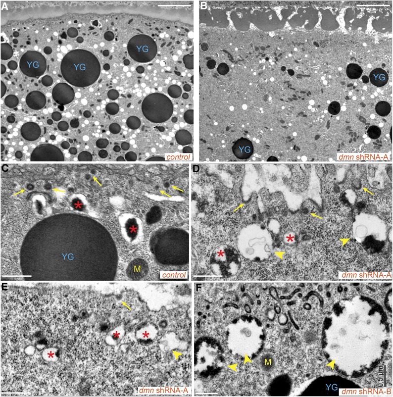
Figure 7.
Ultrastructural analysis of Dmn-depleted oocytes. (A and B) Control egg chambers (A) and egg chambers expressing dmn shRNA-A (B) were fixed and processed for electron microscopy. Bar, 5 μm. (C–F) High-magnification views of control egg chambers (C) and egg chambers expressing dmn shRNA-A (D and E) or dmn shRNA-B (F). Bar, 500 nm. “YG” indicates condensed yolk granules. “M” indicates mitochondria. Arrows indicate coated pits and coated vesicles. Arrowheads indicate endocytic intermediate structures containing some yolk and intraluminal vesicles. Asterisks indicate endocytic vesicles with partially condensed yolk proteins. In most of these vesicles, the yolk proteins remained attached to the membrane.

