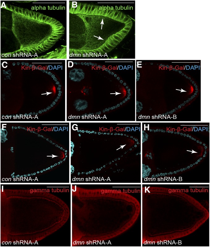
Figure 8.
Microtubule polarity in Dmn-depleted oocytes. (A and B) Egg chambers expressing a control shRNA (A) and dmn shRNA-A (B) were fixed and processed for immunofluorescence using an antibody against α-tubluin (green). Arrows indicate reduced α-tubulin staining along the cortex of Dmn-depleted egg chambers. (C–E) Egg chambers expressing a control shRNA (C), dmn shRNA-A (D), or dmn shRNA-B (E) along with the kinesin β-gal reporter were fixed and processed for immunofluorescence using an antibody against β-galactosidase (red). The oocytes were also counterstained with DAPI to reveal nuclei (cyan). Kinesin β-gal is a marker for microtubule plus-ends. Stage 9 egg chambers are depicted. The arrow indicates posterior Kinesin β-gal. (F–H) Stage 10 egg chambers from strains expressing a control shRNA (F), dmn shRNA-A (G), or dmn shRNA-B (H) along with the kinesin β-gal reporter were fixed and processed for immunofluorescence using an antibody against β-gal (red). The oocytes were also counterstained with DAPI to reveal nuclei (cyan). The arrow indicates posterior Kinesin β-gal. (I–K) Egg chambers expressing a control shRNA (I), dmn shRNA-A (J), or dmn shRNA-B (K) were fixed and processed for immunofluorescence using an antibody against γ-tubulin (red). γ-Tubulin is a marker for microtubule minus-ends. Bar, 50 μm.

