Figure 9.
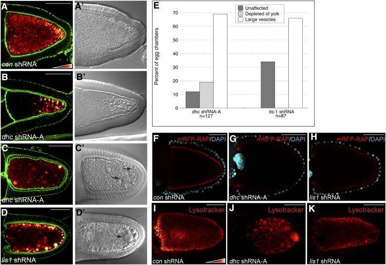
Dynein and Lis1 are required for endocytosis. (A–D) The following strains were fixed and stained to reveal the actin cytoskeleton: con shRNA (A), dhc shRNA-A (B and C), and lis1 shRNA (D). The shRNAs were expressed using a maternal α-tubulin-Gal4 driver. Autofluorescent yolk particles are displayed using a color-coded range indicator. DIC images of these egg chambers are shown in A′, B′, C′, and D′. The efficacy of Lis1 depletion is shown in Figure S1I. (E) Quantification of endocytic phenotypes. Egg chambers from the indicated genotypes were scored for the presence of yolk and large vesicular structures. The percentage of each phenotype observed and the number of egg chambers counted for each genotype are indicated. (F–H) Egg chambers expressing a control shRNA (F), dhc shRNA-A (G), or lis1 shRNA (H) were processed for mRFP-RAP endocytosis (red). The egg chambers were incubated with mRFP-RAP for 30 min. They were then fixed and stained with DAPI to reveal nuclei (cyan). (I–K) Egg chambers expressing a control shRNA (I), dhc shRNA-A (J), or lis1 shRNA (K) were processed for Lysotracker staining. The Lysotracker signal is shown using a color-coded range indicator. Bar, 50 μm.

