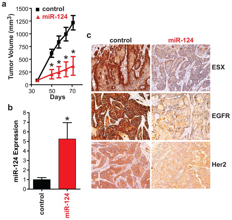
Figure 3. Restoration of miR-124 suppresses the tumorigenicity of HNSCC in vivo.
(a) Tumor growth. SCC15/miR-control or SCC15/miR-124 cells were injected subcutaneously to the flanks of the nude mice. Tumors were measured using a caliper and tumor volumes were calculated. Data is presented as mean ± SEM. *p<0.01, n=7. (b) Intratumoral miR-124 expression. SCC15/miR-control and SCC15/miR-124 tumors were resected and total RNA was isolated. miR-124 expression was measured using qPCR. Data is presented as mean ± SEM. *p<0.01. (c) Intratumoral ESX, EGFR and Her2 levels. ESX, EGFR and Her2 levels was assessed by standard immunohistochemistry. A representative image is shown for the SCC15/miR-control and SCC15/miR-124 tumors.

