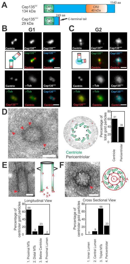
Figure 1. Cep135 isoforms exhibit unique localization patterns.
(A) Cep135 protein isoforms. CR, conserved region. Red, C-terminal tail is divergent from Cep135full. (B) Localization of Centrin (α-Centrin, red), γ-tubulin (α-γ-Tub; green), Cep135full (α-Cep135full, green), and Cep135mini (Alexa488-α-Cep135mini, cyan and red) during G1. Cep135full and Cep135mini localize to the proximal end of G1 centrioles. Scale bar, 1.0 μm. (C) During G2, Cep135mini localizes to centrioles and PCM. Scale bar, 1.0 μm. (D) Immuno-EM localization of Cep135mini to the centriolar microtubules and the PCM (n=152 gold particles for 33 centrioles). (E and F) Cep135mini localizes to the proximal centriole triplet microtubules. (n=47 gold particles). (D–F) The relative distribution of gold particles was quantified for localization to the centrioles and the PCM, centriolar longitudinal sections, and centriolar cross sections, respectively. Red arrowheads denote gold localization. Scale bar, 100 nm.

