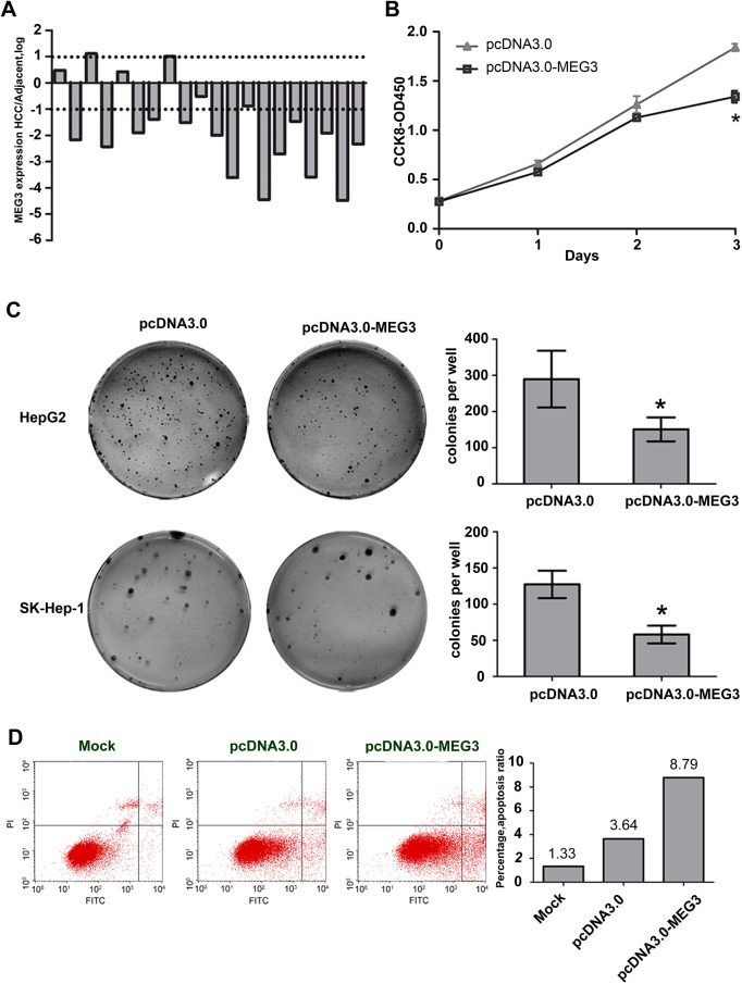
Fig 4. Overexpression of MEG3 inhibited growth of hepatoma cells and induces cell apoptosis.
(A) MEG3 was remarkably reduced in HCC tissues. Reduced and undetected samples account for 82% and 13% of the total samples. Total RNA was extracted from human HCC and adjacent nontumorous tissues with Trizol. Expression of lncRNA MEG3 was examined by qRT-PCR and normalized to GAPDH. The bars represent the ratio of MEG3 between HCC and adjacent nontumorous tissues (log scale). (B) The effects of MEG3 on viability of cells were detected by using Cell Counting Kit–8. The value are means of three independent experiments ±SD, * P<0.05. (C) The effects of MEG3 on cell growth were detected by using colony formation assay in HepG2 and SK-Hep–1. The value are means of three independent experiments ±SD, * P<0.05. (D) Ectopic expression of MEG3 induced cell apoptosis. Cell apoptosis distribution of HepG2 cells transfected with pcDNA3.0 or pcDNA3.0-MEG3 after serum-starved for 24 hours. The apoptotic rates of cells were detected by flow cytometry. This experiment was independently performed at least three times and the change tendency is the same. One of the results was shown in Figure.

