
Fig 1. Western Blot Analysis of BMPRII Protein in the Brain.
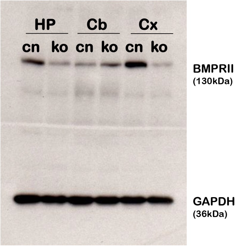
Western blot analysis of tissues from the hippocampus (HP), Cerebellum (Cb), and cortex (Cx) in BMPRII flox/flox controls (cn) and BMPRII flox/flox; CaMKIIα-Cre (ko) mice. At 2 months old, fbΔBMPRII mutant mice show a great reduction of BMPRII protein in the hippocampus and cortex, but show similar levels of BMPRII protein in the cerebellum.