Fig. 4.
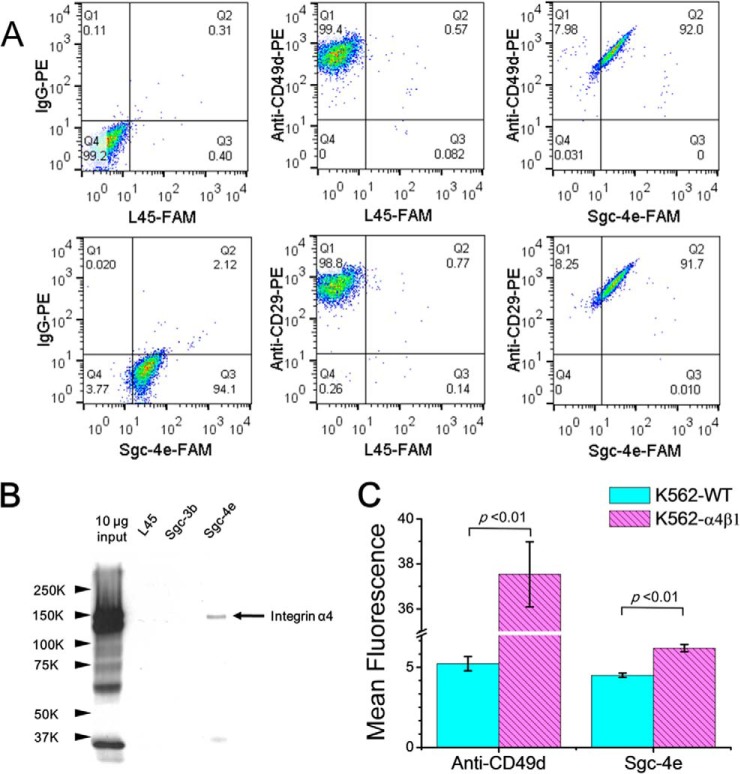
Validation of protein targets of aptamer Sgc-4e. (A) Flow cytometry assay results of Jurkat E6–1 cells stained with Sgc-4e-FAM and anti-CD49d-PE or anti-CD29-PE. FAM-labeled random DNA (L45-FAM) and IgG-PE, which do not bind to cells, were used as negative control. Sgc-4e-FAM and anti-CD49d-PE or anti-CD29-PE can bind simultaneously to Jurkat E6–1 cells. (B) Western blot analysis of proteins that were pulled down by random DNA sequence (L45), control aptamer (Sgc-3b) and aptamer Sgc-4e, respectively. (C) Flow cytometry assay results showed that K562 cells with stable expression of α4β1 display stronger binding to Anti-CD49d or Sgc-4e than wild-type K562 cells.

