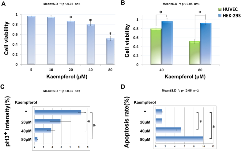
Figure 4. Kaempferol selectively inhibited mammalian endothelial cells and affected cell proliferation and apoptosis process.
(A) inhibition of HUVECs viability by kaempferol in a dose-dependent manner. (B) kaempferol inhibited HUVECs more effectively than HEK-293 cells. HUVECs and HEK-293 cells were treated with different concentrations of kaempferol for 24 hours. Cell viability was quantified by MTT assay. (C,D) statistics of pH3/DAPI and TUNEL/DAPI double staining in HUVECs after 48 hours treatment with different concentration of kaempferol. The pattern is representative of three similar results. *P < 0.05 versus control group. Scale bar, 100 μm.

