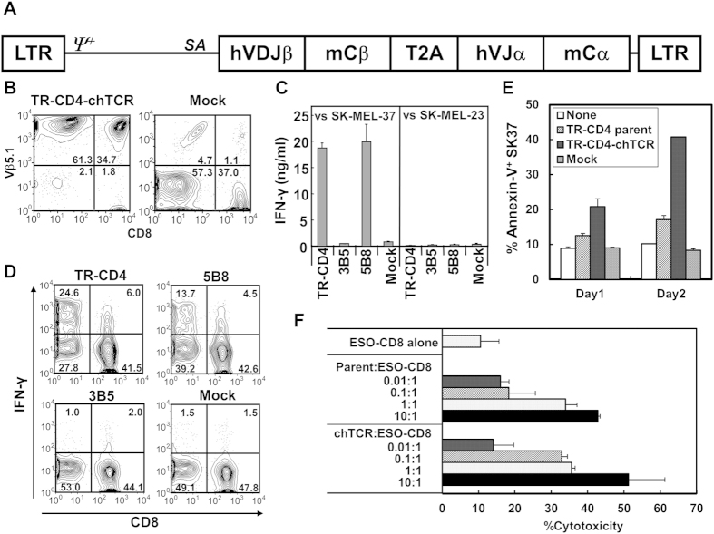
Figure 6. Generation of MHC-II-restricted tumor-recognizing T cells by retroviral TCR gene-engineering.
(A) Schematic representation of chTCR expression vector. TCR expression cassettes were constructed for TR-CD4, 3B5, and 5B8 CD4+ T cell clones and inserted into pMS3 retroviral plasmid vector. LTR: long terminal repeats; ψ+: extended packaging signal; SA: Splice acceptor site from the first exon-intron junction of human elongation factor-1α; hVDJβ: human TCR β chain variable-diverse-joining regions; mCβ: murine TCR β chain constant region; T2A: SGSG-linker connected to the T2A translational skipping sequence; hVJα: human TCR α chain variable-joining regions; mCα: murine TCR α chain constant region. (B) Expression of TR-CD4-chTCR. Preactivated human PBMC were infected twice with retroviruses-transducing TR-CD4-chTCR or no vector control (Mock) and 2 days after the second infection, cell surface expression of Vβ5.1 was investigated by flow-cytometry. (C) TR-CD4, 3B5, or 5B8-chTCR-transduced PBMC or uninfected (Mock) PBMC were cocultured with DP4+NY-ESO-1+ SK-MEL-37 or DP4+NY-ESO-1− SK-MEL-23 for 24 hours. IFN-γ level in the supernatant was measured by ELISA. Error bars represent the standard deviation from 2 independently infected PBMC from a healthy individual. (D) chTCR-transduced or Mock infected PBMC were cocultured with SK-MEL-37 for 6 hours in the presence of Brefeldin A. Expression of IFN-γ was measured by flow-cytometry after staining with cell surface CD8 and CD4 (not shown) and intracellular IFN-γ. Percentage of IFN-γ+ cells after coculture with NY-ESO-1− cell lines was less than 2% in both CD4+ and CD8+ T cells. (E) Induction of apoptosis on SK37 by TCR-transduced CD4+ T cells. CD8-depleted PBMC were preactivated and transduced with TR-CD4-chTCR as described above. SK37 (1 × 10⁵ cells) were cocultured with or without parental TR-CD4, TR-CD4-chTCR-transduced CD4 or Mock-transduced CD4 at 2 × 10⁵ cells. Apoptotic cell death on SK37 was determined by staining with anti-annexin V antibody at day 1 and day 2. (F) SK37 was co-cultured with ESO-CD8 at 1:2 ratio in the presence or absence of indicated numbers of parental TR-CD4 or TR-CD4-chTCR-transduced CD4. Cytotoxicity was evaluated by CFSE-based cytotoxicity assays. All experiments were repeated using PBMC from 2 different healthy individuals with similar results.

