. 2015 Aug 29;4:e08647. doi: 10.7554/eLife.08647

Figure 3. KPNB1 mediates PERs/CRYs-regulated transcription of E-box dependent clock genes.

(A) HEK293T cells were transiently transfected with Per1-Luc reporter construct alone or cotransfected with plasmids expressing CLOCK, BMAL1, PER1, PER2, CRY1, CRY2, in the presence of control (siCTL: black), CSE1L (siCSE1L: light green), KPNB1 (siKPNB1; gray red), and TNPO1 siRNA (siTNPO1: violet) as indicated. After 24 hr, the cells were lysed and Per1 promoter-driven luciferase activities were measured and normalized with pRL-TK activity. Results of one representative experiment of three independent experiments are shown. (B) Schematic diagram of the human PER1 promoter and primers used for ChIP assay. (C, D) ChIP assay of PER1 or PER2 binding to the E box in hPER1. Control (siCTL: black) and KPNB1 siRNA (siKPNB1; gray red)-treated cells (U2 OS) were subjected to ChIP assays using anti-PER1 (αPER1) or anti-PER2 (αPER2) antibody. ChIP DNA samples were quantified by quantitative real-time RT-PCR. The data presented are the means ± S.E. of triplicate samples (**p < 0.005, *p < 0.05, by Student's t-test). (E) Quantitative real-time RT-PCR analysis of expression of endogenous PER1, CRY1, DBP, REVERBβ, and BMAL1 mRNAs in control (siCTL: black), KPNB1-depleted (siKPNB1; gray red) or overexpressed (KPNB1OV; blue green) cells (U2 OS). The data presented are the means ± S.E. of triplicate samples (**p < 0.005, *p < 0.05, by Student's t-test).

DOI: http://dx.doi.org/10.7554/eLife.08647.008

Figure 3.

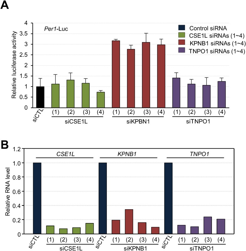
Figure 3—figure supplement 1. Depletion of KPNB1 reduces repressional activity of PER and CRY proteins.

Figure 3—figure supplement 1.

(A) Effects of knockdown of TNPO1, KPNB1, CSE1L on Per1 promoter activity. HEK293T cells were transiently transfected with Per1-Luc reporter construct in the presence of control siRNA (siCTL: dark blue) or four siRNAs targeting each of CSE1L (siCSE1L 1–4: light green), KPNB1(siKPNB1 1–4; gray red), and TNPO1 siRNA (siTNPO1 1–4: violet) as indicated. After 24 hr, cells were lysed and Per1 promoter-driven luciferase activities were measured and normalized with pRL-TK activity. Results of one representative experiment of three independent experiments are shown. (B) Quantitative RT-PCR analysis of knockdown efficiencies of four siRNAs targeting each of CSE1L (siCSE1L 1–4: light green), KPNB1 (siKPNB1 1–4; gray red), and TNPO1 siRNA (siTNPO1 1–4: violet) introduced in HEK293T cells as indicated.
Figure 3—figure supplement 2. Deletion of KPNA2, importin α binding domain is not required for regulatory function of KPNB1 in clock gene transcription.

Figure 3—figure supplement 2.

(A) Schematic illustration of full-length (1–876) and mutant (1–771) KPNB1 engineered with a deletion of the importin-α binding domain on the C-terminus. (B) Deletion of the importin-α binding domain does not affect the down-regulation of Per1 promoter activity by KPNB1. HEK293T cells were transiently transfected with Per1-Luc reporter construct alone (left) or cotransfected with plasmids expressing full-length and mutant KPNB1 (KPNB1-WT, KPNB1-[1–771]) as indicated combination. After 24 hr, cells were subjected to lysis and Per1 promoter-driven luciferase activities were measured and normalized with pRL-TK activity. The data presented are the means ± S.E. of triplicate samples. (C) Deletion of the importin-α binding domain does not affect the down-regulation of CLOCK-BMAL1-mediated Per1 transcription by KPNB1. HEK293T cells were transiently transfected with Per1-Luc reporter construct alone (left) or cotransfected with plasmids expressing CLOCK, BMAL1, full-length or importin-α binding domain deficient mutant of KPNB1 (KPNB1-WT, KPNB1-[1–771]) as indicated combination. Similar transcriptional analysis was performed, as in (B).