Figure 3.

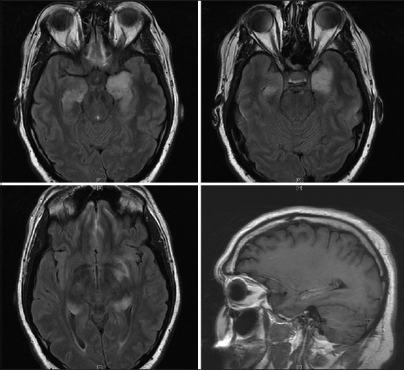
Axial T2 flair magnetic resonance images demonstrating hyperintensity in the bilateral mesial temporal lobes left greater than right, as well as contrast enhancement, in right mesial temporal lobe on sagittal section

Axial T2 flair magnetic resonance images demonstrating hyperintensity in the bilateral mesial temporal lobes left greater than right, as well as contrast enhancement, in right mesial temporal lobe on sagittal section