
Figure 4.
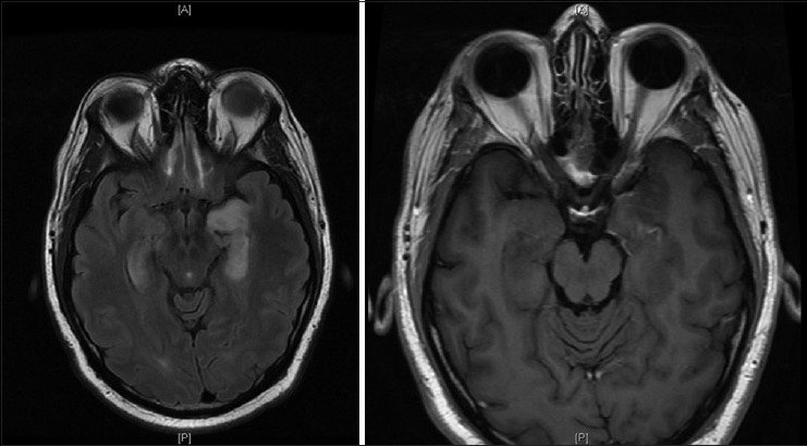
Axial magnetic resonance T2 flair image on the left and postcontrast T1 image on the right showing interval improvement in enhancement pattern with stable hyperintensity of bilateral mesial temporal lobes
Official websites use .gov
A
.gov website belongs to an official
government organization in the United States.
Secure .gov websites use HTTPS
A lock (
) or https:// means you've safely
connected to the .gov website. Share sensitive
information only on official, secure websites.
Axial magnetic resonance T2 flair image on the left and postcontrast T1 image on the right showing interval improvement in enhancement pattern with stable hyperintensity of bilateral mesial temporal lobes