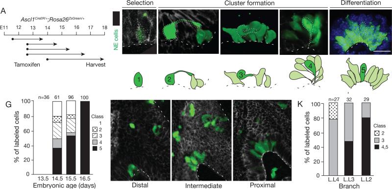
Figure 5. Sparse labeling of progenitors reveals a series of NEB intermediates.
(A) Labeling scheme. NE progenitors were permanently labeled with ZsGreen by tamoxifen injection of Ascl1CreER/+;Rosa26ZsGreen/+ mice at indicated ages (dots), then allowed to develop for times indicated (arrows).
(B-F) Micrographs and schematics of five classes of NE intermediates (green) labeled as above and co-stained for E-cadherin to outline epithelial cells. A representative cell of each class is outlined (dots) in micrograph and highlighted in dark green in schematic; other labeled NE cells are shown in light green in schematic. Note long (>10 μm) apical cell extension (class 2), fibroblast-like morphology and no basement membrane contact (class 3), and thin cellular extensions that converge at basement membrane (class 4). In late embryo (~E17) through adult, all NE cells contact basement membrane (class 5). Bar, 10 μm. See also Figures S5A,B and Table S3.
(G) Quantification of different classes of NEB intermediates labeled as above and harvested 48 hours after tamoxifen injection at ages indicated. The changing abundance of intermediates supports their ordering in a developmental series. n, number of intermediates scored in 2 lungs at each stage.
(H-K) Confocal images (sagittal sections; H-J) and quantification (K) of NEB intermediates labeled as above at base of bronchi L.L4, L.L3, and L.L2 in same lung. L.L4 is most distal (immature) and L.L2 is most proximal (mature) position. The different abundance of intermediates along proximal-distal axis supports their ordering in a developmental series. n, number of intermediates scored in 2 embryos. Bar, 10 μm.

