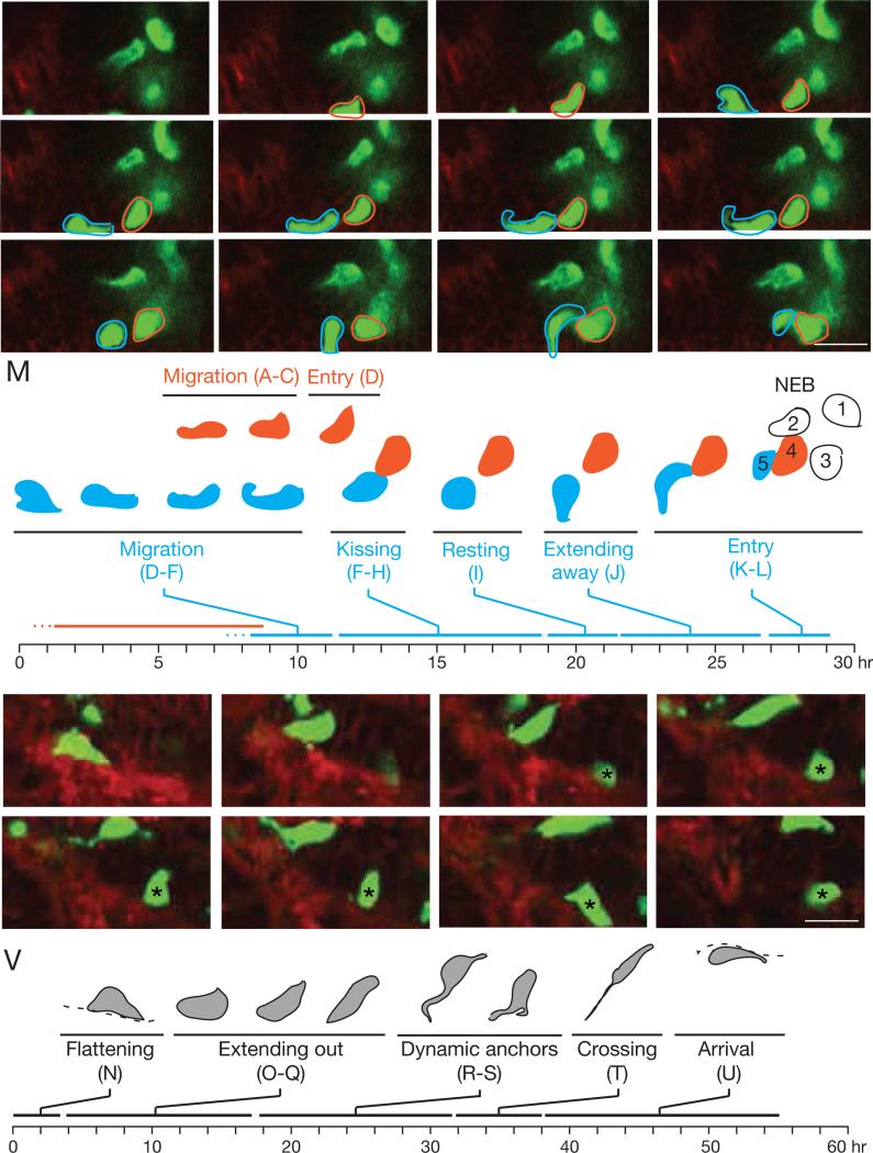
Figure 6. Live imaging of migrating NE progenitors in lung slice culture.
(A-L) NE cell migration and entry into NEB. Selected frames from 26 hour time-lapse confocal microscopy (Movie S1) of migrating NE cells in E15 mouse lung slice culture from Asc1CreER/+;Rosa26ZsGreen/mTmG mouse induced with tamoxifen at E13 to label NE cells with ZsGreen and mGFP (green) and other cells with mTomato (red). NE cells 1-3 at developing NEB site are joined by cell 4 (red outline), which migrates into region (B, C; at ~1.6 μm/hr) and directly enters cluster (D). Cell 5 (blue outline) migrates into same region (D; ~1.6 μm/hr) but soon changes direction and extends toward cell 4 (E). Cell 4 reciprocates (F) and the cells contact briefly (G, “kissing”) and retract (H), repeating kissing sequence 4 times in ~5 hours. Over the next 5 hours, cell 5 remains stationary (I, “resting”) before extending backward (J, “extending away”) and then diving forward (K, “entry”) to join NEB cluster (L).
(M) Timeline of above changes in cell 4 (red) and 5 (blue).
(N-U) NE cell crossing bronchial tube. Selected frames from another region of same culture. Initially, NE cell flattens along basement membrane (N, “flattening”) then re-orients toward lumen (O-Q, “extending out”). Over next ~14 hours, its contact with basement changes dynamically (R,S, “dynamic anchors”), including formation of a second extension that contacts basement membrane >10 μm away (S). Just before crossing, anchor is a very fine projection with “beaded” appearance (T) that fragments as cell finally crosses (~2.5 μm/hr) into epithelium of opposing side of tube (crossing, T) to reach destination (U, arrival). Frame U was selected from a z-plane 8 μm deeper than panels N-T to more clearly visualize cell after arrival. Dashed line, basement membrane. Bar, 10 μm. See also Movies S1, S2.
(V) Timeline of above changes.

