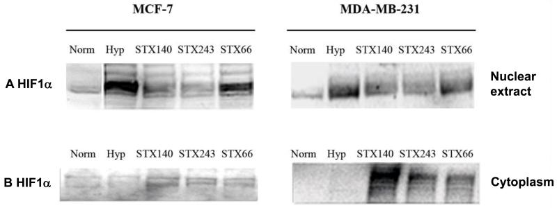
Figure 2.
Hypoxia-inducible Factor 1α (HIF1α) protein expression in MCF-7 and MDA-MB-231 cells. MCF-7 and MDA-MB-231 cells were treated with 0.5 μM STX140, STX243 or STX66 under normoxia for 18 h and then under hypoxia (1% O2, 5% CO2) for 6 h. Equal amount of each nuclear extract (A) or cytoplasmic fraction (B) was resolved by Sodium Dodecyl Sulfate-Polyacrylamide Gel Electrophoresis and blotted for HIF1α. The immunoblot shown is representative of three separate experiments and was quantified using Kodak 1D software. A: MCF-7 nuclear extracts, lane 1: control normoxia (100% intensity); 2: control hypoxia (241%); 3: STX140 (162%*); 4: STX243 (105%*); 5: STX66 (216%). MDA-MB-231 nuclear extracts, lane 1: control normoxia (100% intensity); 2: control, hypoxia (147%); 3: STX140 (128%*); 4: STX243 (123%*); 5: STX66 (144%). B: MCF-7 cytoplasmic extracts, lane 1: control normoxia (100% intensity); 2: control hypoxia (75%); 3: STX140 (146%*); 4: STX243 (134%*); 5: STX66 (122%*). MDA-MB-231 cytoplasmic extracts, lane 1: control normoxia (100% intensity); 2: control hypoxia (67%); 3: STX140 (259%*); 4: STX243 (173%*); 5: STX66 (162%*). *p<0.05, compared to control.

