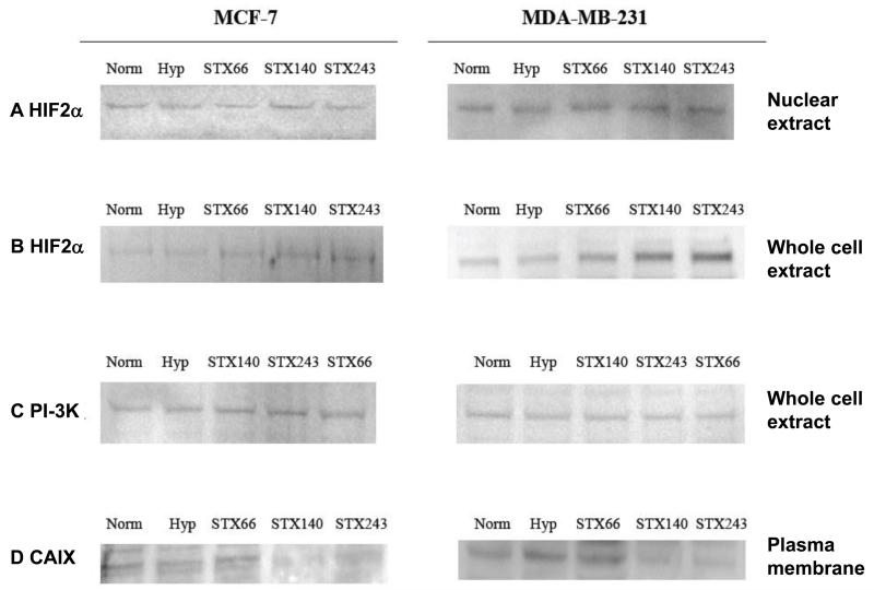
Figure 5.
Hypoxia-inducible Factor 2α (HIF2α), Phosphatidylinositol-3 Kinase (PI-3K) and Carbonic Anhydrase IX (CAIX) protein expression in MCF-7 and MDA-MB-231 cells. MCF-7 and MDA-MB-231 cells were treated with 0.5 μM STX140, STX243 or STX66 under normoxia for 18 h and then under hypoxia (1% O2, 5% CO2) for 6 h. Nuclear extracts (A) and total protein extracts (B) were prepared and analyzed by Sodium Dodecyl Sulfate-Polyacrylamide Gel Electrophoresis and immunoblotting with the antibody to HIF2α or PI-3K (C). Cells were given an additional 48 h incubation without compound and plasma membrane fractions were prepared and analyzed by SDS-PAGE and immunoblotting with the antibody to CAIX (D). A: MCF-7 nuclear extracts, lane 1: control normoxia (100% intensity), 2: control hypoxia (99%); 3: STX66 (101%); 4: STX140 (101%); 5: STX243 (102%). MDA-MB-231 nuclear extracts, lane 1: control normoxia (100% intensity); 2: control hypoxia (101%); 3: STX66 (102%); 4: STX140 (104%); 5: STX243 (103%). B: MCF-7 total protein extracts; lane 1: control normoxia (100% intensity); 2: control hypoxia (102%); 3: STX66 (108%); 4: STX140 (119%); 5: STX243 (123%). MDA-MB-231 total protein extracts, lane 1: control normoxia (100% intensity); 2: control hypoxia (99%); 3: STX66 (108%); 4: STX140 (121%*); 5: STX243 (134%*). C: MCF-7 total protein extracts, lane 1: control normoxia (100% intensity); 2: control hypoxia (100.2%); 3: STX140 (100.3%); 4: STX243 (99.6%); 5: STX66 (100.3%). MDA-MB-231 total protein extracts, lane 1: control normoxia (100% intensity); 2: control hypoxia (99.8%); 3: STX140 (99.2%); 4: STX243 (98.8%); 5: STX66 (99.3%). D: MCF-7 plasma membrane fraction, lane 1: control normoxia (100% intensity); 2: control hypoxia (101.3%); 3:STX66 (103%); 4: STX140 (61.2%*); 5: STX243 (74.2%*). MDA-MB-231 plasma membrane fraction; 1: control (100% intensity); 2: control hypoxia (104%); 3: STX66 (99.23%); 4: STX140 (67.3%*); 5: STX243 (67.4%*). *p<0.05, compared to control.

