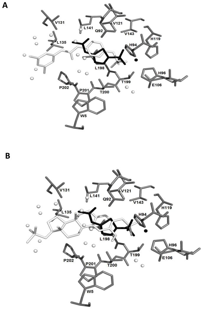
Figure 6.
S4 (A) and STX140 (B) docked into the 3IAI ‘A’ chain crystal structure of CAIX. Amino acid residues are shown in grey and are labelled. The crystal structure acetazolamide is shown in black. The black sphere is a zinc ion. The docked ligands are shown in white. The white spheres are the oxygen atoms of water molecules.

