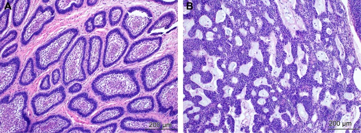
Figure 1.

Histopathology of ameloblastoma. (A) The follicular pattern with islands of odontogenic epithelium within fibrous stroma. The epithelium consists of peripheral palisading cells showing reverse polarization and central loosely arranged cells resembling the stellate reticulum. H&E staining (×100). (B) The plexiform pattern with anastomosing strands of basal cells, delicate stroma, and inconspicuous stellate reticulum. H&E staining (×100).
