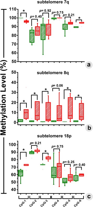
Fig. 3.

The changes in methylation level (%) were quantitatively determined at each of the CpG sites by pyrosequencing. Significant changes in the percentage of methylation were observed in two out of six CpGs in Chr. 7q (a), five out of six CpGs in Chr. 8q (b), and two out of six CpG sites in Chr. 18p (c). Methylation in glioma and control patients is presented with green and red colored box-plots, respectively
