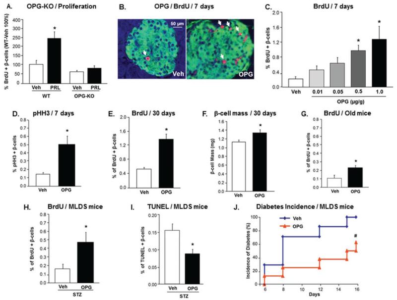
Figure 1. OPG is required for PRL-induced rodent β-cell proliferation, and enhances rodent β-cell replication in young, aged, and STZ-treated mice.
(A) Percent BrdU-positive β-cells in male OPG KO and WT mice infused with Veh or PRL for 7 days. β-cell proliferation in WT-Veh mice is depicted as 100% (n=4-5 mice/group); *p<0.05 vs other three groups; (See also Figure S1). (B) Representative images of pancreatic sections stained for insulin (green) and BrdU (red) from mice injected daily for 7 days with vehicle (Veh) and 1.0μg mOPG-Fc/g body weight. Arrows indicate BrdU- and insulin-positive cells. (C) Percentage of BrdU-positive β-cells in 10-week old mice injected daily for 7 days with Veh or different doses of mOPG-Fc; (n=5-10 mice/group, >1000 β-cells counted/mouse; See also Figure S2). (D) Percentage of pHH3-positive β-cells in mice treated as in (B). (E) Percentage of BrdU-positive β-cells in mice injected every alternate day with Veh or 1.0μg/g mOPG-Fc/g for 30 days; (n=5 mice/group; See also Figure S3). (F) β-cell mass in mice treated as in (E); *p=0.05 vs Veh. (G) Percent BrdU-positive β-cells in one-year old mice injected daily with Veh or 1.0μg/g mOPG-Fc for 7 days (n=6-7 mice/group). (H) Percent BrdU-positive β-cells in MLDS mice injected daily with Veh or 1.0μg/g mOPG-Fc for 16 days (n=7-8 mice/group). (I) Percent TUNEL-positive β-cells in mice treated as in (H). (J) Diabetes incidence, defined as blood glucose >250mg/dl, in mice treated as in (H); #p<0.05 vs Veh by chi-square test. All values are presented as mean ± SEM. *p<0.05 vs Veh, except where specified otherwise.

