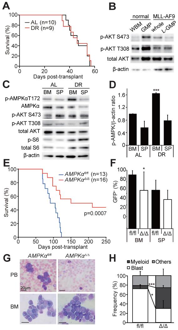
Figure 1. AMPK is activated in AML cells upon DR and promotes leukemogenesis.
(A) Secondary recipients of 1,000 MLL-AF9-induced AML cells were either fed ad libitum (AL, n=10) or subjected to dietary restriction (DR, n=9). DR did not extend the survival of AML recipient mice. (B) Immunoblotting was performed on freshly isolated whole bone marrow cells (WBM) and GMPs from non-leukemic mice, and whole AML cells (sorted GFP+ cells) and L-GMPs from leukemic mice, all fed AL. PI3K pathway, as determined by phosphorylation of Akt, was not activated in AML cells compared to normal progenitors. (C) Immunoblotting of freshly isolated AML cells from AL or DR mice revealed that AMPK was highly activated in the bone marrow (BM), but not the spleens (SP), of DR mice. (D) Quantification of the results shown in (C). p-AMPK signals were normalized to the signals from β-actin. (E) Survival of mice after transplanting MLL-AF9 transduced AMPKαfl/fl (n=13) or AMPKαΔ/Δ cells (n=16, three independent experiments). (F) Frequencies of GFP+ AML cells in the bone marrow or the spleens of leukemic mice shown in (E). (G) Wright-giemsa staining of peripheral blood (PB) or the bone marrow (BM) samples from AML mice revealed fewer blasts in the recipients of AMPKαΔ/Δ AML. (H) Quantification of the cell types in the bone marrow. In all figures, data represent mean±standard deviation; *, p<0.05; **, p<0.005; and ***, p<0.0005 by Student’s t-test, except for comparison of the survival curves in which the significance was accessed by a log-rank test. See also Figure S1.

