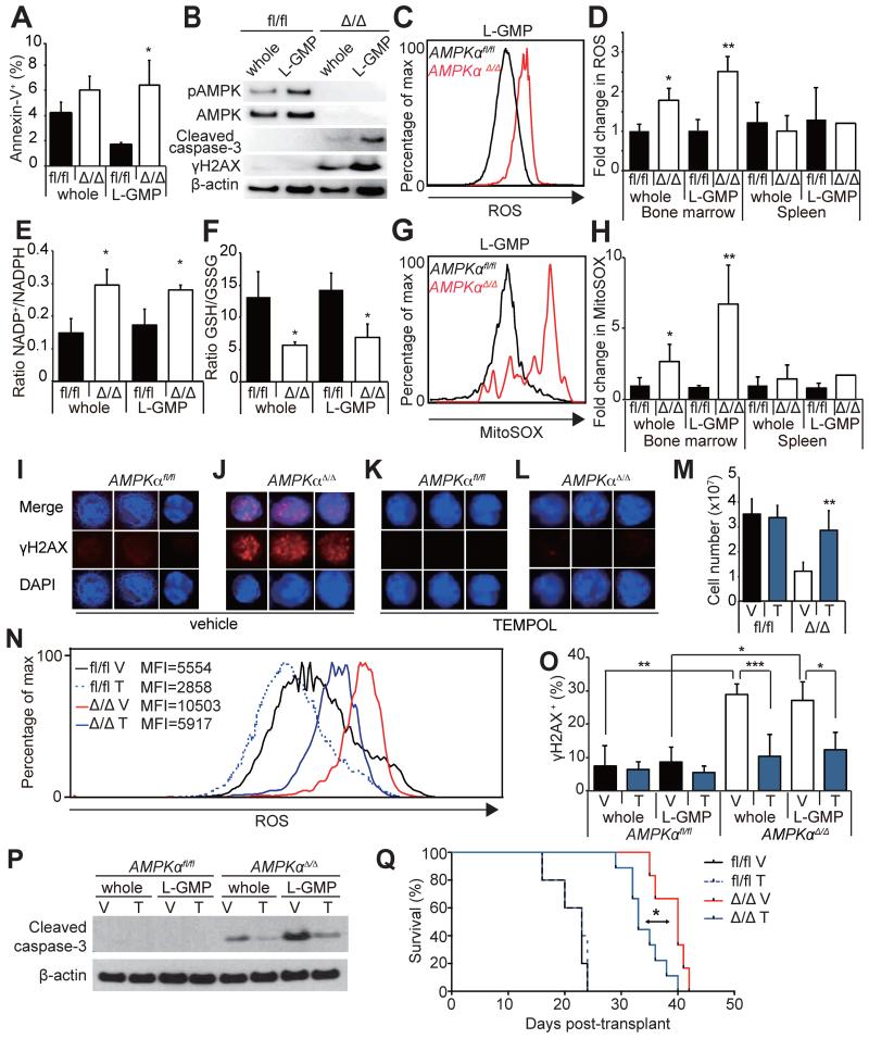
Figure 4. AMPK deletion induces oxidative stress and DNA damage in LICs.
(A) AMPK deletion significantly increased the frequencies of Annexin-V+ L-GMPs in the bone marrow (n=3). (B) Western blotting of AMPKαfl/fl and AMPKαΔ/Δ AML cells and L-GMPs from the bone marrow revealed increased cleaved caspase-3 consistent with the increased cell death, as well as increased γH2AX indicating increased DNA damage. (C and D) AMPK deletion increased the levels of ROS in whole AML cells and L-GMPs in the bone marrow but not in the spleens (n=5). (E and F) Deletion of AMPK from L-GMPs and whole AML cells from the bone marrow increased the NADP+/NADPH ratio and reduced the GSH/GSSG ratio consistent with the increased oxidative stress (n=3). (G and H) AMPK deletion also increased the levels of mitochondrial superoxide (as indicated by the MitoSOX staining) in whole AML cells and L-GMPs in the bone marrow but not in the spleens (n=5). (I-L) Immunofluorescence staining of freshly isolated bone marrow L-GMPs with anti-γH2AX antibodies revealed that AMPK deletion (J) increased DNA damage, which was significantly suppressed by treating the mice with an antioxidant TEMPOL (L). Scale bars indicate 5 μm. Quantification of I-L is shown in O (V: vehicle, T: TEMPOL, n=5). (M) TEMPOL treatment increased the numbers of AMPKαΔ/Δ AML cells in the bone marrow, and reduced ROS levels in bone marrow L-GMPs (N). MFI indicates the mean fluorescence intensity of each sample. (P) Western blotting of AMPKαfl/fl and AMPKαΔ/Δ AML cells and L-GMPs isolated from the bone marrow of mice subjected to TEMPOL or vehicle treatment revealed that the levels of cleaved caspase-3 in AMPKαΔ/Δ AML cells and L-GMPs were partially reduced by TEMPOL treatment. (Q) 104 GFP+ AML cells from secondary recipients were transplanted and recipients treated with either control vehicle or TEMPOL. TEMPOL treatment slightly but significantly accelerated the onset of leukemogenesis by AMPKαΔ/Δ AML cells.

