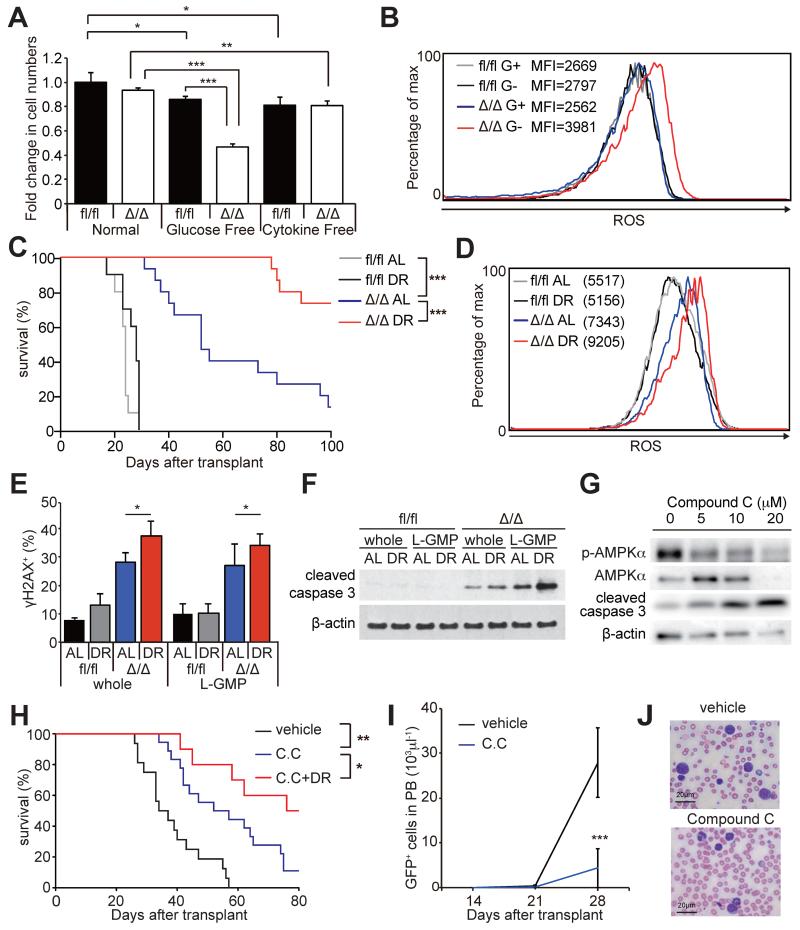
Figure 7. AMPK inhibition sensitized AML to dietary restriction.
(A) In vitro glucose deprivation, but not cytokine deprivation, significantly reduced survival of AMPKαΔ/Δ AML cells during a 24-hour culture (n=4). (B) Representative histograms showing increased ROS levels in AMPKαΔ/Δ AML cells after glucose deprivation in vitro. G+ and G− indicates cultured with or without glucose, respectively. (C) Secondary recipients of 104 AMPKαfl/fl (n=10 per group) and AMPKαΔ/Δ AML cells (n=15 per group) were either fed AL or subjected to DR and their survival monitored. DR did not extend the survival of AMPKαfl/fl AML recipients, but did significantly extend the survival of AMPKαΔ/Δ AML recipients. (D) Representative histograms showing that DR further increased the higher ROS levels of AMPKαΔ/Δ L-GMPs (n=3). (E) The frequencies of whole AML cells and L-GMPs positive for γH2AX examined by immunofluorescence assay. (F) Immunoblotting assay using AMPKαfl/fl and AMPKαΔ/Δ whole AML cells and L-GMPs isolated from the bone marrow of the recipient mice subjected to AL or DR. (G) Immunoblotting assay of primary AML cells treated with compound C. (H) Secondary recipient mice of 103 MLL-AF9 AML cells were fed AL or subjected to DR after transplantation. 7 days after transplantation, mice were treated with 4 mg/kg of compound C (C.C) or control vehicle. Compound C treatment significantly extended the survival, and this was further extended when combined with DR (vehicle; n=16, C.C; n=18, C.C+DR; n=10). (I and J) Compound C treatment significantly reduced the total numbers of GFP+ AML cells (I) and the frequencies of blasts (J) in peripheral blood (vehicle; n=5, C.C; n=8). Scale bars indicate 20 μm. See also Figure S6.

