
Figure 1. In vivo bone formation in rat calvarial defects implanted with collagen scaffolds.
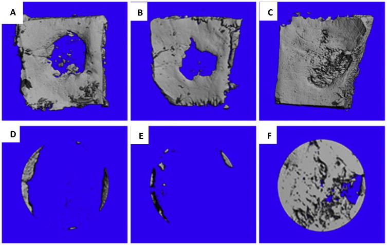
Representative microCT scans showing the level of regenerated bone tissue in calvarial defects 4 weeks after implantation with empty defects (no collagen scaffolds) (A, D), empty scaffolds (collagen scaffolds alone) (B, E,) and PEI-pPDGF-B complex-loaded scaffolds(collagen scaffold with complex) (C, F). Images D, E, F are close up images of the defects and shows the rate of bone regeneration[69]. Reprinted from Biomaterials, 35, Satheesh Elangovan, Sheetal R. D'Mello, Liu Hong, Ryan D. Ross, Chantal Allamargot, Deborah V. Dawson, Clark M. Stanford, Georgia K. Johnson, D. Rick Sumner, Aliasger K. Salem, The enhancement of bone regeneration by gene activated matrix encoding for platelet derived growth factor, 737-747, Copyright 2014, with permission from Elsevier.