
Figure 10. Effect of scaffolds on bone regeneration.
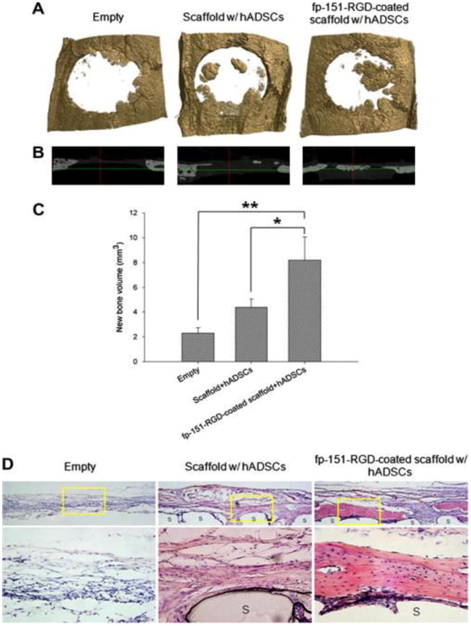
Calvarial defects in rats were implanted with various (indicated) PCL/PLGA blended scaffolds made using fused deposition modeling. (A) μCT scan 3-D and (B) X-ray 3-D axis images of rat calvaria and (C) quantified new bone volume after 8 weeks of implantation. (A) represent the calvarial defect margin. Values and error bars represent the means of quintuple samples and standard deviations with statistical significance (*p < 0.05 and **p < 0.01). (D) Histological analysis of in vivo bone regeneration 8 weeks after implantation. hADSC = human adipocyte-derived stem cells; fp-151-RGD = a genetically redesigned hybrid mussel adhesive proteins[108]. Reprinted from Acta Biomaterialia, 8, Jung Min Hong, Bum Jin Kim, Jin-Hyung Shim, Kyung Shin Kang, Ki-Joo Kim, Jong Won Rhie, Hyung Joon Cha, Dong-Woo Cho, Enhancement of bone regeneration through facile surface functionalization of solid freeform fabrication-based three-dimensional scaffolds using mussel adhesive proteins, 2578-2586, Copyright 2012, with permission from Elsevier