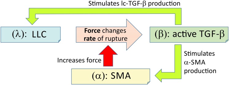
Fig 3. A diagram showing how the proteins in our mechanosensing model interact.
The three key markers of the signalling loop are: λ (the concentration of LLC), β (the concentration of active TGF-β) and α (the concentration of SMA). The rate of spontaneous breaking of λ, k m(α, κ), is a non-linear function of the pulling force F applied by the cell, Eq (1).

