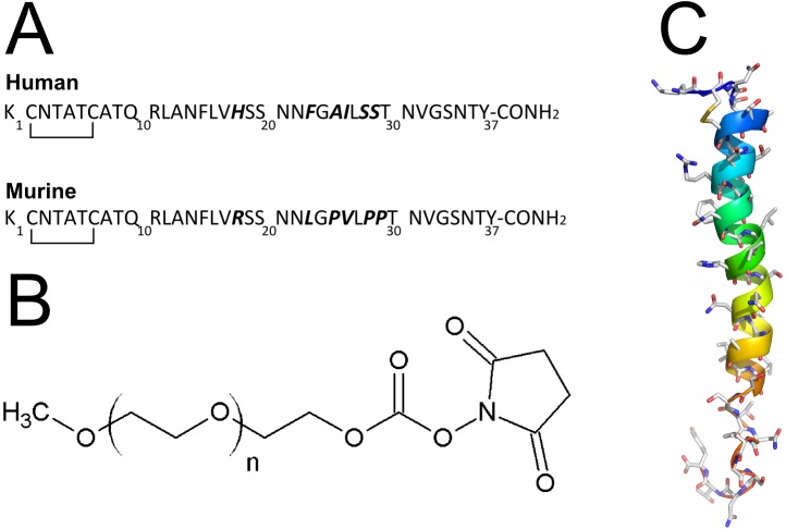
Fig 1. Amylin and PEG structures.
a) Aminoacid sequence of the human and murine amylin. In bold are the different aminoacid. Notice the similarity in sequence from aminoacid 1 to 17. The N-terminus is the Lys1, which comprises the α- and ε-aminogroup, the two unique primary aminogroups in amylin targeted for PEGylation. b) Chemical structure of the methoxyl PEG N-hydroxylsuccinimide (NHS) carbonate. c) Structure of the human amylin. Human amylin (from NMR structure, PDB ID 2KB8) is represented in ribbons colored from aminoacid 1 (blue) to 37 (red). Notice the two aminogroups at the top left side of the representation.

