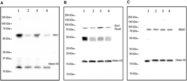
Fig 4. Western Blot analysis of differentially expressed genes.
(A) Chk1, (B) Emi1/Fbxo5, (C) Mybl2 protein were detected by western blot analysis. Histone H3 served as the normalization control. Murine mouse muscle cells were cultured for 48 h in growth medium (lane 1), differentiation medium (lane 2), or differentiation medium supplemented with TNF-α (lane 3) or IGF1 (lane 4), respectively. (B) The specificity of the double band between 40 and 50 kDa was confirmed by peptide competition of the Emi/Fbxo5 antibody epitope (S4 Fig).

