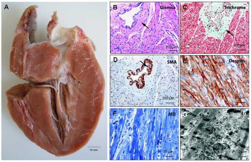
Figure 3.
A: Pathological examination of the explanted heart at age 36 weeks showing massive biventricular hypertrophy, thickened endocardium, and small ventricular cavities. B-D: Microscopic analysis depicting marked perivascular fibrosis and profound wall thickening of an arteriole (arrows). (e, g) Desmin staining, methylene blue stained semi-thin section (MB), and electron microscopy demonstrating disarray of myofibers and Z-bands.

