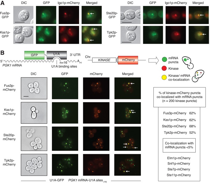
Fig 4. Co-localization of Fus3p, Kss1p, Ste20p, and Tpk2p with mRNPs.
A) The RNP component Igo1p was visualized as a carboxy-terminal mCherry fusion generated by integration of a mCherry cassette at the 3’-end of IGO1. GFP chimeras were constructed as integrated in-frame fusions to the 3’-ends of the indicated kinase genes. Cells were examined after three days of incubation with shaking in liquid cultures of minimal medium with normal levels of ammonium sulfate. Arrowheads indicate foci with the given kinase and Igo1p. Scale bar, 3 μm. Fluorescent protein fusions to the kinases Elm1p, Snf1p, Ste7p, and Ste11p did not localize significantly as puncta under identical conditions (S3A Fig), and were not tested further for co-localization with Igo1p. B) RNPs were visualized as foci using PGK1 modified to contain 16 binding sites for U1A-GFP in its 3’-UTR. GFP-tagged RNA was analyzed for co-localization with kinase-mCherry fusions generated by integration of sequence encoding mCherry as an in-frame fusion to the 3’-end of the targeted kinase gene. Kinase localization was observed after 15 minutes of glucose stress in SC–Leu–Ura media lacking glucose. Arrowheads indicate kinase-mCherry puncta co-localized with GFP-tagged RNA foci. Quantification of puncta is provided.

