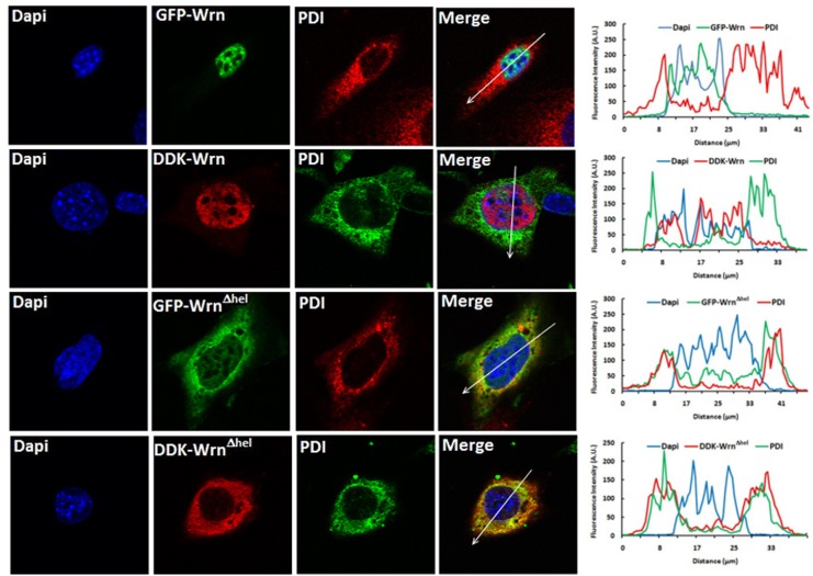
Fig 7. Example of co-localization of the WrnΔhel mutant protein with the ER organelles by immunofluorescence study.
Images in the first row represent the localization of the GFP-Wrn and PDI in Wrn Δhel/Δhel MEFs. Images in the second row represent the localization of the DDK-Wrn and PDI in Wrn Δhel/Δhel MEFs. Images in the third row represent the localization of the GFP-WrnΔhel mutant protein and PDI in Wrn Δhel/Δhel MEFs. Images in the fourth row represent the localization of the DDK-WrnΔhel mutant protein and PDI in Wrn Δhel/Δhel MEFs. The graph at the end of each row represents the intensity of the fluorescence along the arrow in the merge image.

