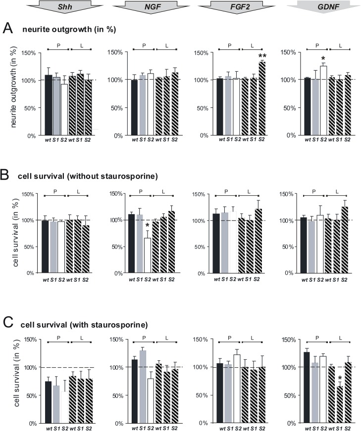
Fig 2. Sulf1 and Sulf2 differentially modulate NGF, FGF2 and GDNF signaling pathways and thereby influence neurite outgrowth and cell survival of cerebellar granule cells.
(A) Cerebellar microexplant cultures from wildtype (wt), Sulf1 (S1) and Sulf2 (S2) deficient mice were plated onto glass cover slips coated with PLL (P, filled bars) or a combination of PLL and laminin (L, hatched bars). Following incubation for 24 h at 37°C with 10 ng/ml Shh, 50 ng/ml FGF 2, 10 ng/ml GDNF or 50 ng/ml NGF, explants were fixed and stained. Neurite outgrowth was quantitated by measuring the ten longest neurites of ten aggregates in three independent experiments. Neurite length of explants of each genotype cultured without any additives was set to 100%. Asterisks indicate a statistically significant difference (** p< 0.01, * p< 0.05). (B, C) Cerebellar neurons from wildtype (wt), Sulf1 (S1) and Sulf2 (S2) deficient mice were plated as single cell suspensions onto PLL coated cover slips (P, filled bars) or cover slips coated with a combination of PLL and laminin (L, hatched bars). Following incubation of growth factors (see A), cell death was determined by counting calcein versus propidium iodide positive cells (B). (C) 20 hours after growth factor treatment 500 nM staurosporine was added to the medium and the cells were cultured for additional 4 h. Then cell death was determined (five independent experiments). Cell survival of each genotype without any additives, see data shown in Fig 1C and 1D, was set to 100% (indicated by dashed lines). Thus, the data show the specific effects of the tested growth factors on cell survival separately for each genotype (and not in relation to wildytpe). Asterisks indicate a statistically significant difference (* p< 0.05).

