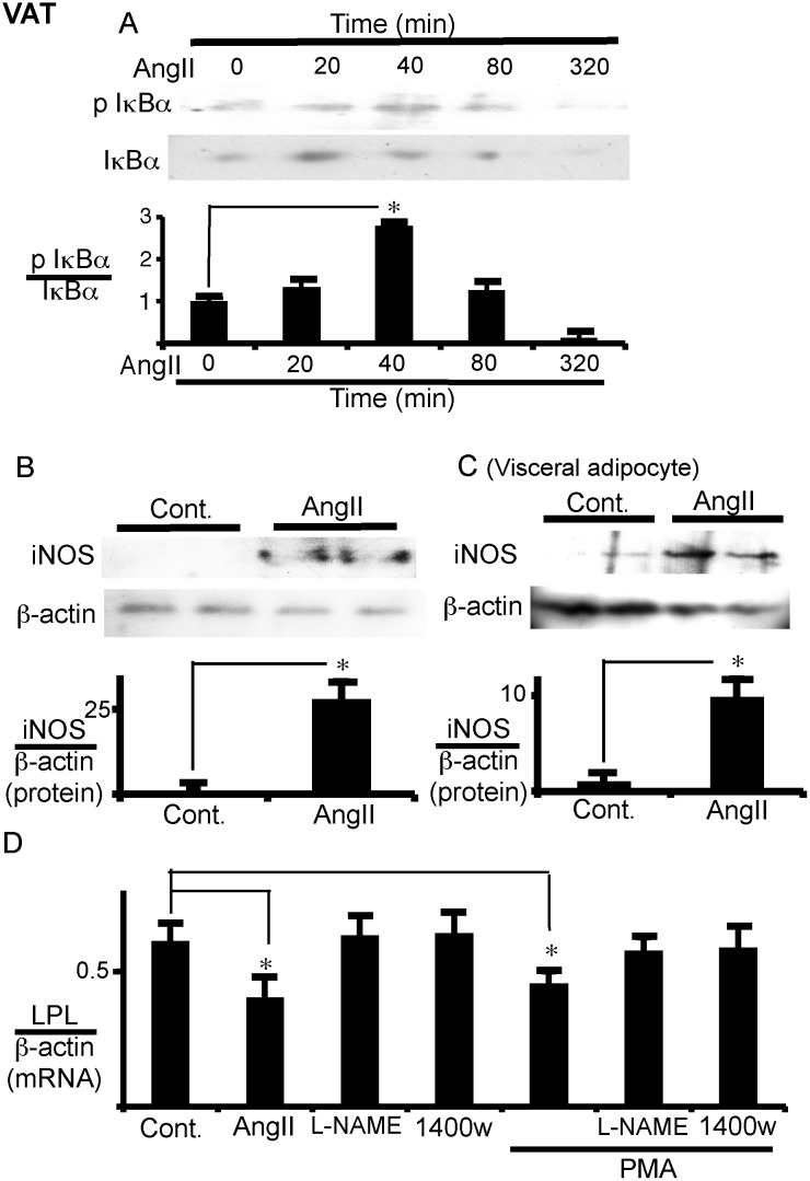
Fig 5. PKC, NFκB, and iNOS mediate the effect of AngII on LPL expression in VAT.
(A) IκBα phosphorylation over time in VAT. VAT was cultured with AngII (1 μM) for the indicated times to measure IκBα phosphorylation with western blotting. The ratio of phospho IκBα to total IκBα was calculated based on densitometric quantification of the bands. (B, C) iNOS expression in VAT and visceral adipocytes. VAT and isolated visceral adipocytes were cultured with or without AngII (1 μM) for 24 h to measure iNOS expression by western blotting. Duplicate samples in each group were processed for western blotting. The ratio of iNOS to β-actin was calculated based on densitometric quantification of the bands (B, VAT; C, visceral adipocytes). (D) PKC is upstream of iNOS in LPL regulation in VAT. VAT was pre-treated with L-NG-nitroarginine Methyl Ester (L-NAME) (1 mM) or 1400w (10 nM) for 1 h prior to phorbol 12-myristate 13-acetate (PMA) (10 nM) addition. After 24 h of AngII (1 μM) or PMA treatment, LPL mRNA expression was measured. The mRNA levels were normalized with β-actin. Each column and bar represents the mean ± SEM for three separate experiments. An asterisk (*) indicates p<0.05 vs. time 0 or without AngII.

