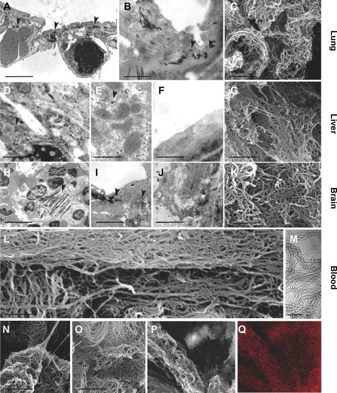
Figure 3.
Ultrastructural analysis of MWCNT aggregates in selected organs.
Notes: TEM and SEM analyses of selected samples of murine tissues showed dark nanoparticle aggregates (arrowheads). Exposed lungs (A–C), liver (D–G), and brain (H–K) in TEM analysis presented dark needle-like structures (arrowheads) with dimensions similar to MWCNTs (A, B, D–F). SEM analysis of the KOH-resistant material confirmed the presence of MWCNTs in the lungs (C), liver (G), brain (K), and blood (L, M) of 5-week MWCNT-exposed animals. These structures were morphologically similar to MWCNT from stock material that has been characterized by SEM before (N) and after (O) KOH treatment, and were not found in unexposed animals. Combination of SEM analysis of MWCNT obtained by KOH corrosion of exposed liver (P) with EDAX (Q) clearly showed co-localization of carbon, indicating that the KOH-resistant material was MWCNT aggregates. Scale bars: (A) 4.5 µm; (B) 1 µm; (C) 1 µm; (D) 1 µm; (E) 1 µm; (F) 0.5 µm; (G) 1 µm; (H) 7.5 µm; (I) 2.7 µm; (J) 2 µm; (K) 0.75 µm; (L) 0.5 µm; (M) 0.1 µm; (N) 1 µm; (O) 1 µm.
Abbreviations: TEM, transmission electron microscopy; SEM, scanning electron microscopy; MWCNT, multi-wall carbon nanotubes; EDAX, energy dispersive X-ray analysis.

