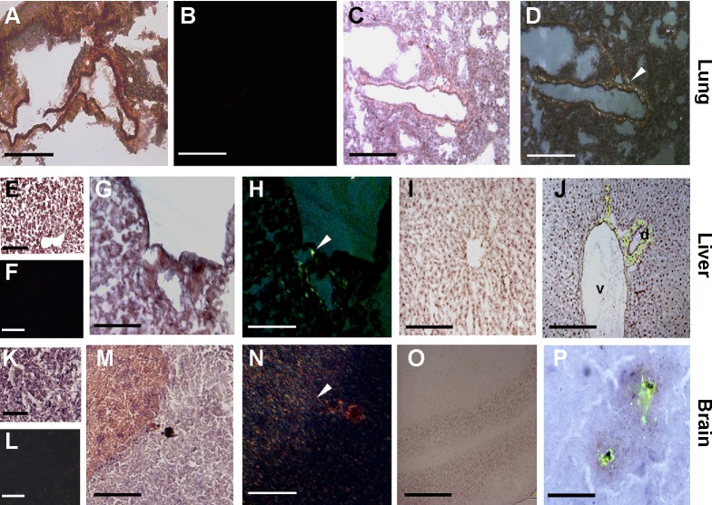
Figure 4.
Detection of amyloid fibril deposits by Congo red and Thioflavin T staining.
Notes: In 5-week MWCNT-exposed mice, birefringence (arrowheads) following Congo red staining indicating amyloid deposition was found in the lung (C, D), liver (G, H), and brain (M, N), while these were not found in the same tissues from unexposed mice (A, B, E, F, K, L). In exposed liver (I, J) Thioflavin T revealed that amyloid fibrils are in association with venules (v) and biliary ducts (d). In the brain (O, P), the amyloid fibrils surround the MWCNT aggregates in what appears to be a nonself response. The scale bar for all photographs =200 µm.
Abbreviation: MWCNT, multi-wall carbon nanotube.

