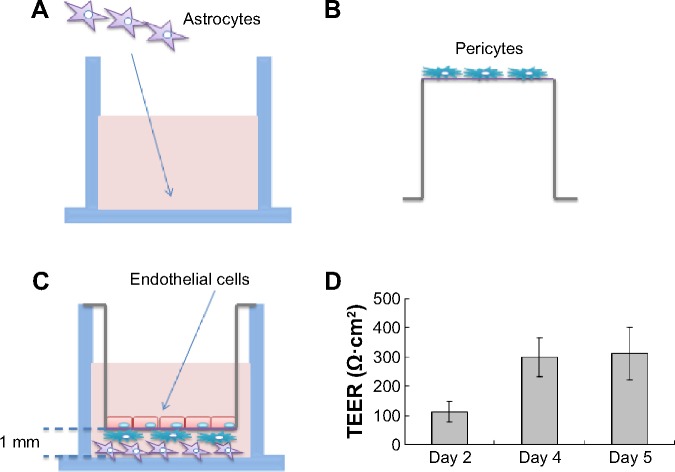
Figure 3.
Schematic drawing of the preparation of the in vitro blood–brain barrier model.
Notes: (A) Rat astrocytes were seeded at the bottom of 12-well plates, while (B) rat brain pericytes were seeded at the against-lumen side of Transwell® membranes of inverted cell culture inserts. After 4 hours, (C) endothelial cells were seeded into the luminal compartment of the inserts having pericytes on the other side and positioned into the 12-well plates containing the astrocytes. (D) TEER (expressed as Ω·cm2) of the blood–brain barrier model, measured on different days. Data are presented as mean ± SD (n=3).
Abbreviations: SD, standard deviation; TEER, transendothelial electrical resistance.

