Figure 6.
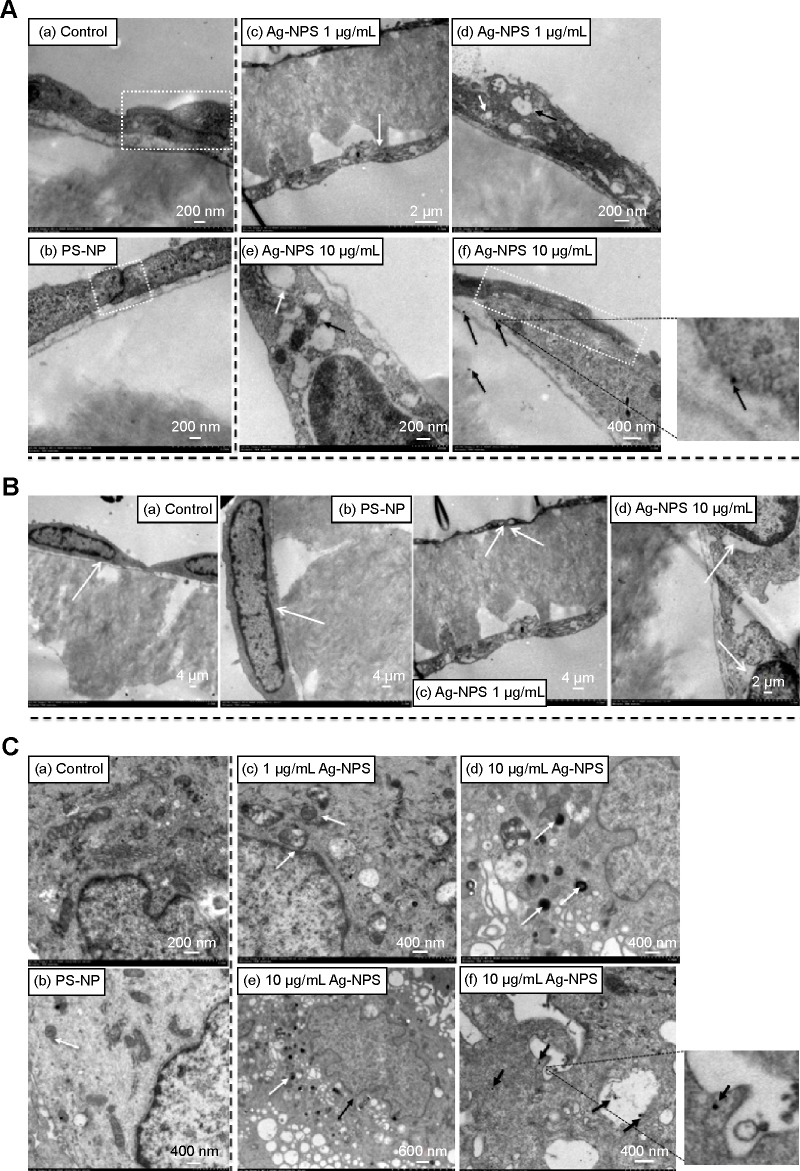
TEM images of primary rat brain (A) microvascular endothelial cells, (B) pericytes, and (C) astrocytes after 24-hour treatments.
Notes: (A) In the control group, endothelial cells form TJs (a, white dash box). In the PS-NP group, a TJ (b, white dash box) between endothelial cells was not disrupted. In the 1 μg/mL Ag-NPS group, the endothelial cells grew nicely on the membrane (c, white arrow) and discontinuous TJs were not observed. Vacuolations (d, white arrow) and ER expansion (d, dark arrow) were observed in the endothelial cells. In the 10 μg/mL Ag-NPS group, mitochondrial shrinkage (e, dark arrow) and ER expansion (e, white arrow) appeared; Discontinuous TJs (f, white dash box) and Ag-NP-like particles as showed in the insert for (f) (f, dark arrows) in endothelial cells were observed. (B) Pericytes grew nicely on the other side of the membrane (white arrow) in the control group (a) and PS-NP group (b). In the 1 μg/mL Ag-NPS group, vacuolations (white arrows) were observed occasionally in pericytes (c). In the 10 μg/mL Ag-NPS group, vacuolations (white arrows) were observed in pericytes (d). (C) In the control group, astrocytes had normal cell morphology (a). In the PS-NP group, mitochondrial shrinkage was observed occasionally (white arrow) (b). In the 1 μg/mL Ag-NPS group, mitochondrial shrinkage was occasionally observed (white arrows) (c). In the 10 μg/mL Ag-NPS group, nuclear atypia (black arrows) and severe mitochondrial shrinkage (white arrows) were observed (d and e); Ag-NP-like particles (showed in the insert for f) were also observed in astrocytes (f, black arrows), as well as in the enlarged picture.
Abbreviations: Ag-NPS, silver nanoparticle suspension; ER, endoplasmic reticulum; PS-NP, polystyrene nanoparticle; TEM, transmission electron microscopy; TJ, tight junction.

