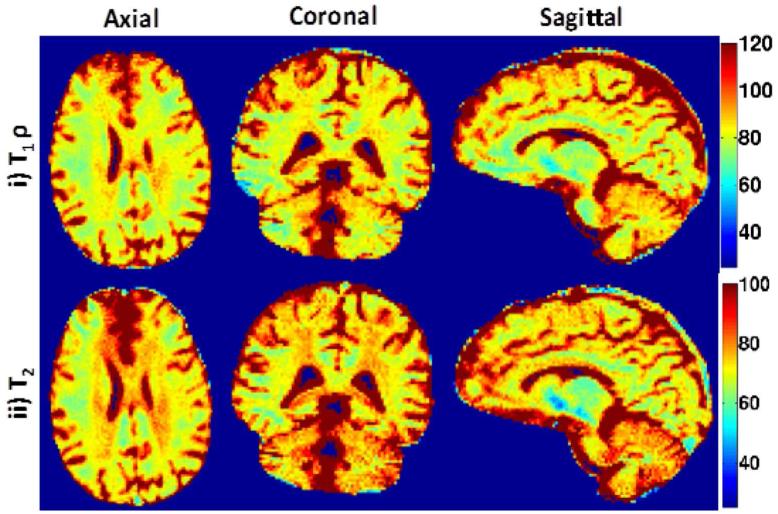
Fig. 10.
Parameter maps for 3D prospective under-sampled data at R=8: Axial, Coronal and Sagittal T1ρ and T2 parameter maps are shown in (i)-(ii). With the acceleration of R=8, the scan time was reduced to 20 min. Note: All 128 slices were processed slice by slice to reconstruct the 3D parameter maps

