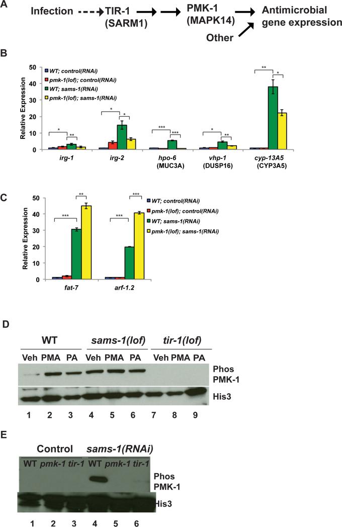
Figure 2. Constitutive activation of innate immune pathway in after sams-1 depletion.
A. Schematic showing p38/PMK-1 mitogen activated protein kinase signaling during response to bacterial infection in C. elegans (Kim, 2014). B. qRT-PCR comparing innate immune gene expression in sams-1(lof) and pmk-1(lof); sams-1(lof) mutants. C. qRT-PCR comparing expression of a lipogenic (fat-7) or other (arf-1.1) gene highly expressed gene in sams-1(lof) and pmk-1(lof); sams-1(lof) mutants. D. Immunoblot of phospho-PMK-1 in vehicle (Veh), phorbol acid treated (PMA) and Pseudomonas aeruginosa (PA) exposed wild type (WT), sams-1(lof) or tir-1(lof) mutants. Histone 3 shows loading. E. Wild type, pmk-1(lof) or tir-1(lof) animals were exposed to control or sams-1(RNAi) and immunoblotted with antibodies to phosph-PMK-1 or Histone 3. Error bars show standard deviation. Results from Student's T test shown by * <0.05, ** <0.01, *** <0.005.

