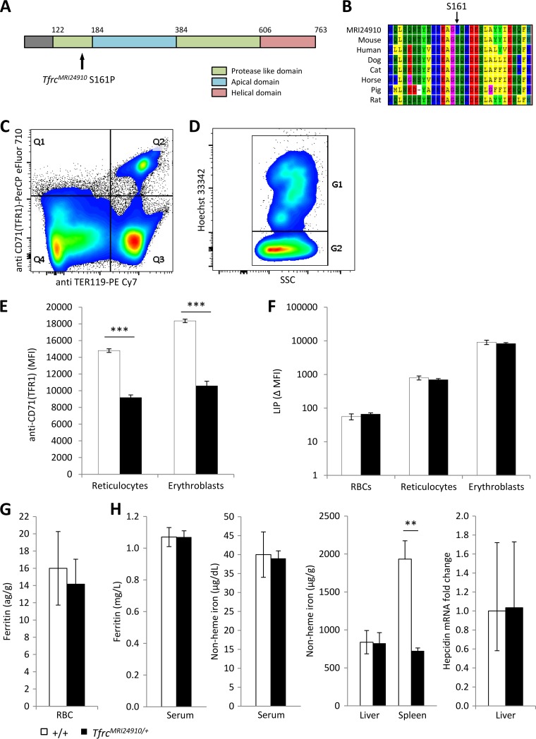
FIG 2.
Identification and characterization of the MRI24910 mutation in Tfrc. (A) An S161P point mutation was located in the protease-like domain of TFR1. (B) The S161 residue is highly conserved, suggesting that it has a critical function in the protein. (C) Splenic erythroblasts and reticulocytes were identified based on positive staining with the markers TER119 and CD71 (TFR1) (Q2), and erythrocytes were identified based on negative CD71 and positive TER119 staining (Q3). (D) From the Q2 gate, erythroblasts (G1) were distinguished from reticulocytes (G2) based on Hoechst staining. (E) The MFI of staining with anti-CD71 (TFR1) was determined for each cell population as a measure of TFR1 surface expression. (F) Labile iron pool (LIP) of each cell population, determined based on the change in CA-AM MFI, with or without the iron chelator L1, according to the method of Prus and Fibach (32). Ferritin contents in erythrocytes (G) and serum (H) were measured by ELISA. (H) Nonheme iron contents in the serum, liver, and spleen were measured colorimetrically, and hepcidin mRNA expression in the liver relative to that of the wild type was determined using the beta-actin gene as a housekeeping gene. Error bars indicate SEM. **, P < 0.01; ***, P < 0.001. Data are representative of one of three experiments with four mice per group for TFR1 MFI. Data are from one experiment with four mice per group for LIP, ferritin, serum iron, and hepcidin measurements and from two experiments with seven or eight mice per group for liver and spleen iron measurements.

