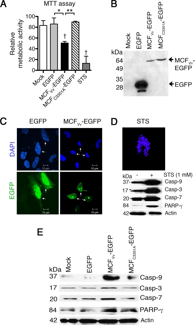
FIG 2.
Ectopic expression of MCFVv inhibits host cell metabolic rate and induces caspase activation. (A) MTT assay on transfected HeLa cells. Data are presented as percent metabolic activity relative to untreated cells. “Mock” indicates cells treated with transfection reagent only. STS was used as a positive control. Results are means and SD for biological triplicates; daggers indicate statistical significance compared to mock-treated cells (P < 0.05), and asterisks show significant difference between the indicated samples (*, P < 0.05; **, P < 0.01). (B) Results of 12% SDS-PAGE showing expression of chimeric proteins by Western blotting using anti-EGFP antibody. (C) Nuclear staining with DAPI of HeLa cells transfected to express the indicated proteins. Arrowheads indicate transfected cells, as shown by detection of green fluorescence (EGFP). (D) DAPI staining of an STS-treated cell and detection of cleaved caspases as a positive control. (E) Representative Western blots of cell lysates from two independent experiments with HeLa cells transfected to express indicated EGFP fusion proteins. Actin is shown as a sample recovery control run in parallel gels along with other proteins.

