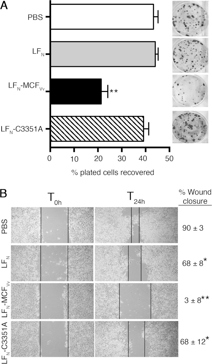
FIG 3.
Direct delivery of MCFVv inhibits host cell proliferation and cell migration. (A) Clonogenic colony formation assay showing percentage of colonies recovered after intoxication with the indicated fusion proteins (72 nM) with PA (168 nM). The cells were seeded at densities of 200 and 400; the recovered colonies were counted, and percent survival was plotted. Quantitative data are reported as percentage of plated cells recovered (means and SD for four biological replicates, each plated twice). **, P < 0.0001 by ANOVA compared to all other samples. The inset shows one representative crystal violet-stained plate for each sample. (B) In vitro wound scratch assay showing photographs of migration patterns and rates of wound closure of HeLa cells within 24 h after intoxication (T24h) with the indicated proteins. T0h indicates the time of wound generation. Values on the right are percentage of the wound healed by cell migration, presented as means ± SD of the percent wound closure. *, P < 0.05, and **, P < 0.0001, by ANOVA followed by multiple comparison to the PBS-treated control.

