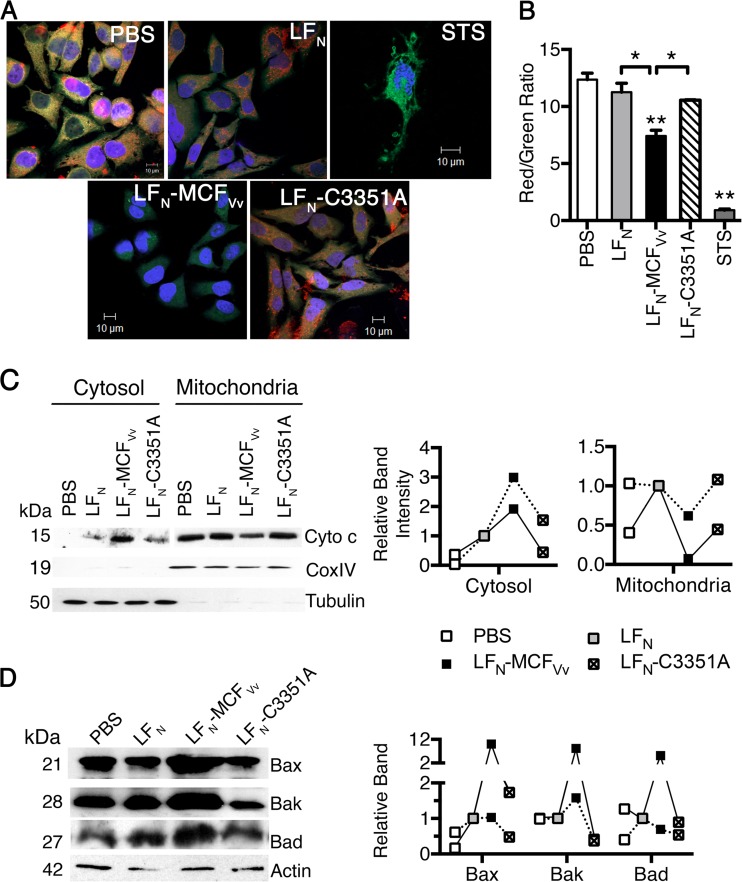
FIG 5.
MCFVv induces mitochondrial damage. (A) JC-1-stained live cells showing red-orange fluorescent aggregates in normal-appearing cells and diffused green fluorescence dispersed throughout the cytoplasm, with minimal red orange fluorescent aggregates in damaged cells. (B) Quantitation of fluorescence intensities from biological triplicate samples similar to those used for panel A was done by measuring the ratio of red (hyperpolarized) to green (depolarized) fluorescence. **, P < 0.01 compared to PBS control; *, P < 0.05 for the indicated comparisons. (C) Cell lysates were fractionated into cytosolic or mitochondria. Anti-cytochrome c antibody was used to probe the level of protein with CoxIV and tubulin proteins used as fractionation markers for mitochondria and cytosol, respectively. (D) Representative Western blots of cell lysates probed for expression levels of the indicated proteins, with actin used as a sample recovery control. On the right is a densitometric quantification of the band intensity for the indicated proteins from two independent experiments (linked by solid or dashed lines) relative to cells intoxicated with unfused LFN protein (gray boxes at 1.0).

