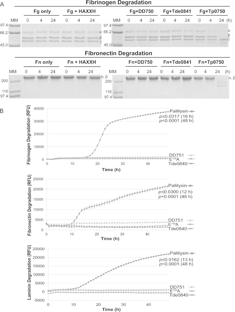
FIG 6.
Tp0750 and pallilysin, but not orthologs from T. denticola and T. phagedenis strain 4A, degrade host proteins. (A) In vitro SDS-PAGE-based assays were performed to compare the host component-degrading capabilities of Tp0750, Tde0841, and DD750. The three recombinant proteins (40 μg each) and the negative-control protein (40 μg), were each incubated with fibrinogen (Fg; 60 μg) or fibronectin (Fn; 30 μg) at 37°C for 24 h. Samples were removed at 0, 4, and 24 h postincubation and analyzed for degradation of the three fibrinogen chains (α, β, and γ) or two comigrating fibronectin chains (α and β) by SDS-PAGE and Coomassie brilliant blue staining (6 μg loaded per lane in the absence of degradation). Numbers to the left of the lanes indicate the size (kDa) of the corresponding molecular mass (MM) markers. All recombinant proteins failed to degrade laminin (data not shown). (B) Fluorescence-based 96-well plate degradation assays were performed to compare the host component-degrading capabilities of pallilysin, Tde0840, and DD751. Recombinant proteins (1 μg per well) were added in triplicate to sterile 96-well plates and incubated with FITC-labeled fibrinogen, fibronectin, or laminin (10 μg per well) at 37 °C for 48 h in the dark. The degree of host component degradation was determined by measuring the increase in relative fluorescence units (RFU) every hour over 48 h using standard fluorescein excitation/emission filters. Average fluorescence intensity readings from triplicate measurements are presented with bars indicating the standard error (SE), and the results are representative of three independent experiments. For statistical analyses, host component degradation by wild-type pallilysin was compared to the highest fluorescence reading from either Tde0840, DD751, or Tp0751 E199A (negative control) using a Student two-tailed t test. Wild-type pallilysin exhibited a statistically significant level of fibrinogen (16 h postincubation; P ≤ 0.0317), fibronectin (12 h postincubation; P ≤ 0.0300), and laminin (13 h postincubation; P ≤ 0.0162) degradation compared to the levels exhibited by Tde0840, DD751, and the negative-control protein.

