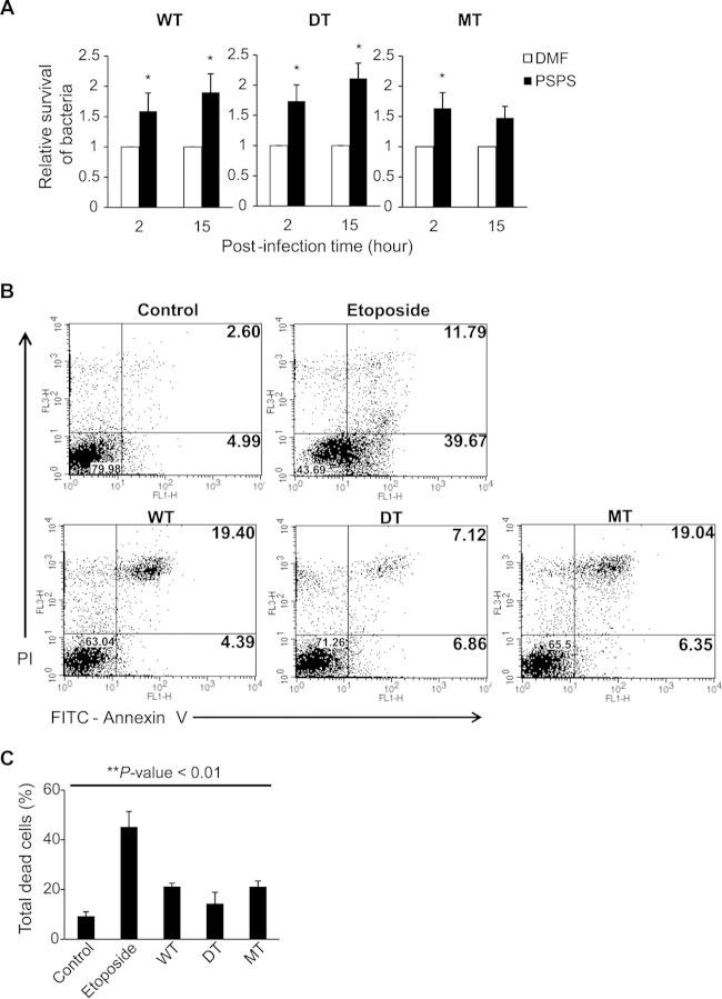
FIG 6.
Effects of MdsABC and phosphatidylserine on bacterial invasion and survival in macrophage-like RAW 264.7 cells. (A) Effect of 50 μM PSPS on the infection of macrophage RAW 264.7 cells by the WT, DT, and mdsABC-overproducing (MT) bacteria. Bacterial invasion and survival were measured from three independent experiments, as described for Fig. 2D, and results are reported as the mean ± SD. Statistically significant differences compared to the WT levels are shown with asterisks: *, P < 0.05. (B) Scatter plots of infected and uninfected macrophages stained with FITC-annexin V (Ann V) and propidium iodide (PI) at 15 h postinfection. Upper panels, scatter plots of uninfected and etoposide-treated RAW 264.7 cells, which represent normal control and apoptosis-induced macrophages, respectively, under the given experimental conditions. Lower panels, scatter plots of RAW 264.7 cells infected by WT, DT, and MT strains with different expression levels of MdsABC. The percentages of viable cells (Ann V− PI−), dead cells (Ann V+ PI+), and apoptotic cells (Ann V+ PI−) were calculated from scatter plots of PS-binding annexin V (Ann V)- and PI-stained dead cells. (C) Percentages of total dead cells (Ann V+ PI+) analyzed using a calibrated FACS flow cytometer. Results from three independent experiments are reported as the mean ± SD. Statistically significant differences compared to the control were analyzed with general one-way ANOVA and paired t tests, with significance set at a P value of <0.01 (**).

