
Figure 5.
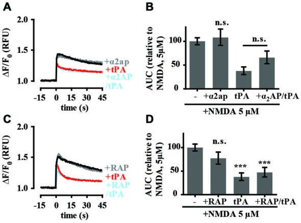
tPA inhibition of NMDA-induced calcium influx is independent of plasmin and lipoprotein receptor-related protein 1 (LRP-1). (A) Hippocampal cultures were preincubated with tPA (40 μg/ml; for 5 min) or α2-antiplasmin (140 nM; for 15 min) and tPA (for 5 min). Baseline Fluo-4 fluorescence was monitored for 15 s prior to the addition of NMDA (5 μM), at time = 0. Fluo-4 fluorescence was monitored for a further 45 s. Raw fluorescence values were converted to ΔF/F0, where F0 is the average fluorescence over the first 15 s of recording prior to addition of agonist (baseline) and ΔF is Fmax−F0. (B) The responses in A were quantitated by measuring the AUC and are presented relative to the AUC for 5 μM NMDA (100%). (C) Hippocampal cultures were preincubated with tPA (40 μg/ml) for 5 min or receptor-associated protein (RAP) (500 nM; for 15 min) and tPA (for 5 min). Baseline Fluo-4 fluorescence was monitored for 15 s prior to the addition of NMDA (5 μM), at time = 0. Fluo-4 fluorescence was monitored for a further 45 s. Raw fluorescence values were converted to ΔF/F0, where F0 is the average fluorescence over the first 15 s of recording prior to addition of agonist (baseline) and ΔF is Fmax−F0. (D) The responses in C were quantitated by measuring the AUC and are presented relative to the AUC for 5 μM NMDA (100%). RFU, Relative Fluorescent Units; n.s, not significant; ***p < 0.001. Error bar, SEM.