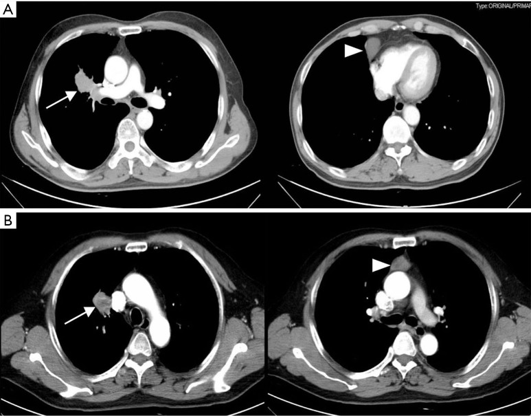
Figure 1.
The chest CT images of two patients. (A) The enhanced chest CT images revealed a 4×2×2 cm3 pulmonary tumor involving both the right main bronchus and pulmonary artery accompanied with a 4×3×2 cm3 cyst in the right anterior mediastinum; (B) the enhanced chest CT images revealed a 3×3×2 cm3 pulmonary tumor adjoined the superior vena cava in the right upper lobe accompanied with a 3×2×2 cm3 thymic tumor in the superior mediastinum. (Arrows indicate the pulmonary tumor; arrowheads indicate the thymic tumor). CT, computed tomography.

