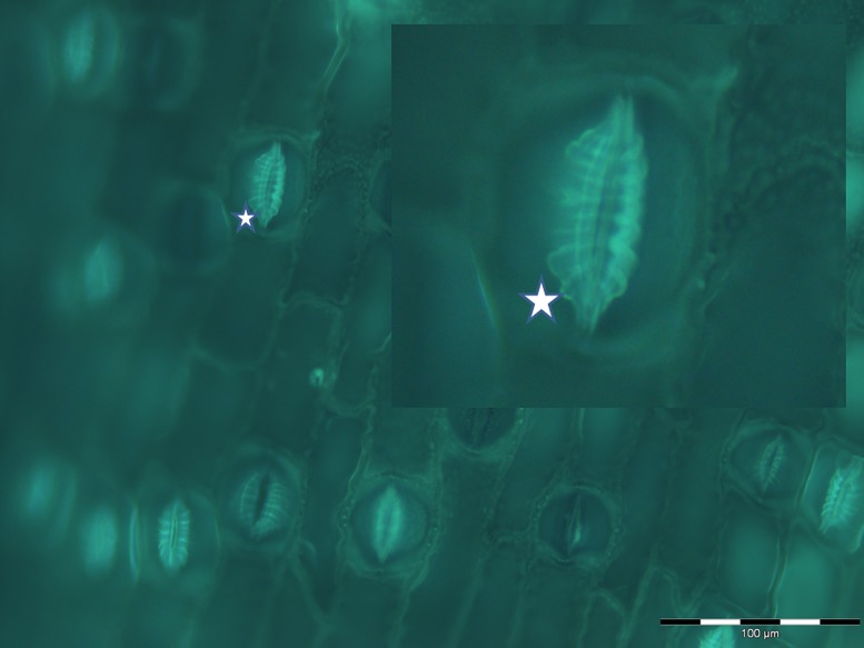
FIGURE 2.
Fluorescent imaging of silica collected following acid and microwave digestion of fern leaf (Asplenium nidus L.) and showing multiple stomata in silicified leaf tissue undergoing differentiation. In particular this image, magnified in the insert, demonstrates how closely silica deposition mimics the deposition of radial fibrillar callose arrays (for example, indicated by star) in stomata in fern (Apostolakos et al., 2009).

