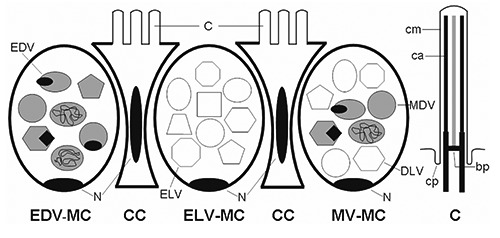
Figure 1.

Schematic diagrams of the esophageal epithelium and cilia. ELV-MC, electronlucent vesicles mucous cell; EDV-MC, electron-dense vesicles mucous cell; MV-MC, mixed vesicles mucous cell; CC, columnar cell; C, cilia; cm, ciliary membrane; ca, ciliary axoneme; cp, ciliary pocket; bp, basal plate; N, nucleus.
